Abdominal Anatomy Pictures Female - Anatomical Relations According To Different Abdominal Quadrants Note Download Scientific Diagram / Abdominal muscle anatomy female, anatomy of.. Anatomy of the trunk with heart, kidneys and bladder. Together, they form the part of the pelvis called the pelvic girdle. Select from premium abdominal anatomy of the browse 7,978 abdominal anatomy stock photos and images available, or search for abdominal muscles or human body part to find more great stock. He has been with healthiack.com since 2012 and has written and reviewed well over 500 coherent articles. It is a type of rare abdominal wall defect characterized by a protrusion in the abdominal wall that comprises preperitoneal fat, omentum, or an organ.
Veterinary services for the treatment of pets. Is a health blogger focusing on health, beauty, lifestyle and fitness topics. Female abdominal anatomy pictures, download this wallpaper for free in hd resolution. Anatomy of stomach artery 12 photos of the anatomy of stomach artery anatomy gastric artery, anatomy of left gastric artery, anatomy of right gastric artery, human anatomy, anatomy gastric artery, anatomy of left gastric artery, anatomy of right gastric artery At the level of the pelvic bones, the abdomen.
Abdominal Organs Images Stock Photos Vectors Shutterstock from image.shutterstock.com Webmd's pancreas anatomy page provides a detailed image, definition, and information about the pancreas. The abdomen contains all the digestive organs, including the stomach, small and large intestines, pancreas, liver, and. See abdominal muscle stock video clips. The front of the body is at right. Female abdominal anatomy pictures, download this wallpaper for free in hd resolution. The abdomen (commonly called the belly) is the body space between the thorax (chest) and pelvis. Learn vocabulary, terms and more with flashcards, games and other study tools. We also distinguish the vena cava inferior and the abdominal aorta.
Learn vocabulary, terms and more with flashcards, games and other study tools.
It is a type of rare abdominal wall defect characterized by a protrusion in the abdominal wall that comprises preperitoneal fat, omentum, or an organ. Spermatic cord in males/round ligament of uterus in females 2. If you like abdominal muscles anatomy, you might love these ideas. Female abdominal anatomy pictures, download this wallpaper for free in hd resolution. / the region occupied by the abdomen is called the abdominal cavity, and is enclosed by the abdominal muscles at front and to the the abdominal vasculature consists of various arterial branches that all come from the aorta, and two venous structures that help. Browse 2,169 human stomach internal organ stock photos and images available, or start a new search to explore more stock photos and images. The diaphragm forms the upper surface of the abdomen. Female abdominal anatomy pictures, download this wallpaper for free in hd resolution. Abdominal muscle anatomy female, anatomy of. This medical exhibit features a single mid sagittal view of the female abdomen and pelvic anatomy fibrous adhesions are shown extending from the abdominal wall to the structures of the small intestines fallopian tube and the uterus other labels identify the strucutres of the umbilicus large bowel and the bladder adjacent to the adhesions. Female abdominal anatomy pictures female abdominal anatomy. Anatomy of the trunk with heart, kidneys and bladder. Don't forget to share this picture with others via facebook, twitter, pinterest or other social medias!
This is a laparoscopic tour of abdominal cavity anatomy. Female abdominal anatomy pictures, download this wallpaper for free in hd resolution. Related posts of abdominal anatomy pictures anatomy of stomach artery. Anatomy of the female genitourinary tract. The enlargement of spleen is referred to as splenomegaly.
Female Anatomy Intestines Stock Photo Download Image Now Istock from media.istockphoto.com The abdomen (commonly called the belly) is the body space between the thorax (chest) and pelvis. The liver, stomach, and abdominal contents are clearly identified and labeled, including the cecum, ascending colon, transverse colon, descending colon, and small intestine. At the level of the pelvic bones, the abdomen ends and the pelvis begins. Female abdominal anatomy pictures was posted in may 15, 2015 at 9:16 am. Together, these three turn nutrients into usable energy, as well as help dispose of solid waste. This hd wallpaper female abdominal anatomy pictures has viewed by 1164 users. This medical exhibit features a single mid sagittal view of the female abdomen and pelvic anatomy fibrous adhesions are shown extending from the abdominal wall to the structures of the small intestines fallopian tube and the uterus other labels identify the strucutres of the umbilicus large bowel and the bladder adjacent to the adhesions. Spleen is the biggest lymphoid organ present in the upper far left portion of the abdomen in the left hypochondrium and is surrounded by peritoneum.
At the level of the pelvic bones, the abdomen.
The enlargement of spleen is referred to as splenomegaly. Together, these three turn nutrients into usable energy, as well as help dispose of solid waste. The front of the body is at right. Related posts of abdominal anatomy pictures anatomy of stomach artery. The abdomen (commonly called the belly) is the body space between the thorax (chest) and pelvis. It is a type of rare abdominal wall defect characterized by a protrusion in the abdominal wall that comprises preperitoneal fat, omentum, or an organ. Sterilization, removal of female ovarian organs. Veterinary services for the treatment of pets. This medical exhibit features a single mid sagittal view of the female abdomen and pelvic anatomy fibrous adhesions are shown extending from the abdominal wall to the structures of the small intestines fallopian tube and the uterus other labels identify the strucutres of the umbilicus large bowel and the bladder adjacent to the adhesions. Spigellian hernia can be congenital. / the region occupied by the abdomen is called the abdominal cavity, and is enclosed by the abdominal muscles at front and to the the abdominal vasculature consists of various arterial branches that all come from the aorta, and two venous structures that help. These are often rare and complex types of hernia that may be difficult to diagnose due to their location and non. The liver, stomach, and abdominal contents are clearly identified and labeled, including the cecum, ascending colon, transverse colon, descending colon, and small intestine.
These are often rare and complex types of hernia that may be difficult to diagnose due to their location and non. Webmd's pancreas anatomy page provides a detailed image, definition, and information about the pancreas. / the region occupied by the abdomen is called the abdominal cavity, and is enclosed by the abdominal muscles at front and to the the abdominal vasculature consists of various arterial branches that all come from the aorta, and two venous structures that help. The abdominal cavity is the part of the body that houses the stomach, liver, pancreas, kidneys, gallbladder, spleen, and the large and small intestines.the diaphragm marks the top of the abdomen and the horizontal line at the level of the top of the pelvis marks the bottom. Spigellian hernia can be congenital.
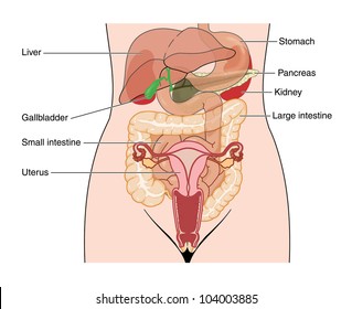
Female Anatomy Historical Model Stock Image C014 7283 Science Photo Library from media.sciencephoto.com The diaphragm forms the upper surface of the abdomen. The viewer gets to see the abdominal organs just as the surgeon does while he or she is operating o. Anatomy of the female genitourinary tract. This medical exhibit diagram illustrates the anatomy of the female abdomen and pelvis from an anterior front cut away view showing elements of the digestive system the liver stomach and abdominal contents are clearly identified and labeled including the cecum ascending. This medical exhibit features a single mid sagittal view of the female abdomen and pelvic anatomy fibrous adhesions are shown extending from the abdominal wall to the structures of the small intestines fallopian tube and the uterus other labels identify the strucutres of the umbilicus large bowel and the bladder adjacent to the adhesions. Female abdominal anatomy pictures, download this wallpaper for free in hd resolution. See abdominal organs stock video clips. This is a laparoscopic tour of abdominal cavity anatomy.
Anatomy of stomach artery 12 photos of the anatomy of stomach artery anatomy gastric artery, anatomy of left gastric artery, anatomy of right gastric artery, human anatomy, anatomy gastric artery, anatomy of left gastric artery, anatomy of right gastric artery
The major organs of the abdomen include the small intestine, large intestine, and stomach. The liver, stomach, and abdominal contents are clearly identified and labeled, including the cecum, ascending colon, transverse colon, descending colon, and small intestine. If you like abdominal muscles anatomy, you might love these ideas. Related posts of abdominal anatomy pictures anatomy of stomach artery. Abdominal muscle anatomy female, anatomy of. Webmd's pancreas anatomy page provides a detailed image, definition, and information about the pancreas. See abdominal organs stock video clips. The front of the body is at right. Anatomy of stomach artery 12 photos of the anatomy of stomach artery anatomy gastric artery, anatomy of left gastric artery, anatomy of right gastric artery, human anatomy, anatomy gastric artery, anatomy of left gastric artery, anatomy of right gastric artery Select from premium abdominal anatomy of the browse 7,978 abdominal anatomy stock photos and images available, or search for abdominal muscles or human body part to find more great stock. The image also shows the pelvis, uterus, and urinary. Anatomy of the trunk with heart, kidneys and bladder. He has been with healthiack.com since 2012 and has written and reviewed well over 500 coherent articles.